Introduction
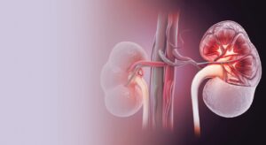
Renal allograft dysfunction, presenting as either acute or gradual loss of kidney function, remains a major complication following transplantation. These conditions often progress silently and are typically detected through an unexplained rise in serum creatinine, sometimes accompanied by proteinuria. Although the underlying mechanisms are multifactorial, cellular- and antibody-mediated rejections and immunosuppressant-related nephrotoxicity are leading contributors to acute and chronic graft injury. This progressive damage ultimately culminates in renal fibrosis and graft loss. Histological assessment via biopsy remains the gold standard for diagnosing the cause of dysfunction, guided by the Banff classification. However, biopsies are invasive and limited by associated risks, while emerging molecular technologies—though promising—are still restricted by their cost and complexity. This highlights the need for advanced, noninvasive biomarkers capable of capturing dynamic changes in graft microarchitecture over time.
Research
MRI-based techniques have gained attention as noninvasive tools to evaluate renal allografts, with diffusion-weighted imaging (DWI) being widely used to characterise renal microstructure. Advanced diffusion approaches such as intravoxel incoherent motion (IVIM) and diffusion tensor imaging (DTI) have shown value in assessing perfusion, fibrosis, and differentiating types of rejection. However, these methods often examine fibrosis and rejection separately, leading to diagnostic gaps when these processes coexist.
Liao and colleagues addressed this challenge by introducing time-dependent diffusion MRI (td-dMRI) to the field of renal transplantation. Previously validated in oncology and hepatology, td-dMRI extends conventional DWI by assessing water diffusion across multiple diffusion times, enabling simultaneous quantification of cellularity, cell diameter, extracellular diffusivity, and intracellular volume fraction. These parameters provide a comprehensive microstructural profile that distinguishes fibrosis from rejection, even when both occur together. Acting as a “virtual biopsy,” td-dMRI offers biologically specific and spatially resolved insights without the need for contrast agents.
In Liao et al.’s study, td-dMRI parameters correlated strongly with clinical and histological disease states. Measures such as cortical cell diameter and intracellular volume fraction differentiated acute from chronic rejection, while medullary cellularity indicated early fibrosis. Diagnostic accuracy was high, with fibrosis and rejection classified at 88.0% and 87.0% accuracy, respectively, outperforming standard clinical assessments with AUC improvements greater than 0.18.
Despite its promise, several limitations remain. The study was single-centre, cross-sectional, and geographically limited, necessitating multicentre and multivendor validation. Technical challenges include the need for harmonisation across MRI systems, standardisation of acquisition protocols, and development of automated processing pipelines. The additional 10 minutes of scan time also raises considerations of patient tolerance and cost-effectiveness. Beyond clinical diagnostics, td-dMRI holds potential for mechanistic research and could be integrated with biomarkers like donor-derived cell-free DNA or combined with complementary MRI techniques—T1 mapping, DTI, ASL—to create a comprehensive multiparametric “virtual biopsy” framework.
Conclusion
Time-dependent diffusion MRI emerges as a powerful and noninvasive imaging tool capable of simultaneously assessing fibrosis and rejection in renal allografts. By bridging the gap between histopathological evaluation and conventional imaging, td-dMRI offers biologically specific microstructural insights that enhance early detection of graft dysfunction. While further standardisation and multicentre validation are required, td-dMRI holds significant potential to guide personalised clinical interventions, improve long-term outcomes, and support multimodal monitoring strategies in both clinical practice and research settings.
References
1. Cornell, L.D. ∙ Helanterä, I.
Exploring microvascular inflammation and the spectrum of antibody-mediated rejection
Am J Transplant. 2025; 25:9-12
2. Callemeyn, J. ∙ Lamarthée, B. ∙ Koenig, A. ∙ et al.
Allorecognition and the spectrum of kidney transplant rejection
Kidney Int. 2022; 101:692-710
3. Virmani, S. ∙ Rao, A. ∙ Menon, M.C.
Allograft tissue under the microscope: only the beginning
Curr Opin Organ Transplant. 2023; 28:126-132
4. Bane, O. ∙ Lewis, S.C. ∙ Lim, R.P. ∙ et al.
Contemporary and emerging MRI strategies for assessing kidney allograft complications: arterial stenosis and parenchymal injury, from the AJR special series on imaging of fibrosis
AJR Am J Roentgenol. 2024; 222, e2329418
5. Caroli, A. ∙ Schneider, M. ∙ Friedli, I. ∙ et al.
Diffusion-weighted magnetic resonance imaging to assess diffuse renal pathology: a systematic review and statement paper
Nephrol Dial Transplant. 2018; 33:ii29-ii40
6. Wang, W. ∙ Yu, Y. ∙ Chen, J. ∙ et al.
Intravoxel incoherent motion diffusion-weighted imaging for predicting kidney allograft function decline: comparison with clinical parameters
Insights Imaging. 2024; 15:49
7. Jiang, B. ∙ Yu, Y. ∙ Wan, J. ∙ et al.
The use of diffusion tensor imaging in the identification of acute rejection and chronic allograft nephropathy after renal transplantation
J Magn Reson Imaging. 2024; 59:2082-2088
8. Liao, Z. ∙ Chen, G. ∙ Liu, K. ∙ et al.
One-stop early noninvasive evaluation of renal allograft rejection and fibrosis: microstructural mapping via time-dependent diffusion MRI
eBioMedicine. 2025; 122, 106034
9. Gore, J.C. ∙ Xu, J. ∙ Colvin, D.C. ∙ et al.
Characterization of tissue structure at varying length scales using temporal diffusion spectroscopy
NMR Biomed. 2010; 23:745-756
10. Mendichovszky, I. ∙ Pullens, P. ∙ Dekkers, I. ∙ et al.
Technical recommendations for clinical translation of renal MRI: a consensus project of the cooperation in science and technology action PARENCHIMA
MAGMA. 2020; 33:131-140