Introduction
(Article introduction authored by Conquest Editorial Team)
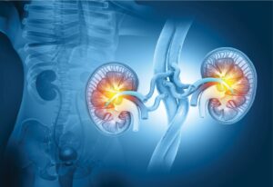
Kidney transplantation stands as the gold standard for treating end-stage renal diseases (ESRDs), offering a life-saving solution for patients. However, the persistent scarcity of donor kidneys has exacerbated the plight of ESRD patients, leaving thousands stranded on transplant waiting lists globally.
Addressing this challenge necessitates innovative approaches to enhance the success rate of kidney transplantation. One promising avenue lies in improving the survival rates of renal grafts, thereby maximizing the utility of available donor organs. Real-time monitoring during the surgical process emerges as a critical factor influencing transplantation outcomes.
Efficient and accurate monitoring techniques can not only ensure the viability of the transplanted kidney but also aid in diagnosing and mitigating potential complications. Unfortunately, existing methods for real-time monitoring of kidney transplantation lack efficiency and precision, underscoring the pressing need for advanced technologies.
In response to this demand, the article introduces a groundbreaking fluorescence technology based on aggregation-induced emission (AIE) active NIR-II nano-contrast agent DIPT-ICF nanoparticles. This novel approach offers comprehensive and real-time monitoring capabilities throughout the entire kidney transplantation procedure. DIPT-ICF exhibits superior photophysical
properties compared to conventional dyes, ensuring bright, stable, and long-lasting fluorescence emission.
Moreover, its extended circulation time enhances its utility in various stages of transplantation surgery, from renal angiography during donor kidney retrieval to assessing reperfusion and diagnosing complications post-implantation.
The study demonstrates that DIPT-ICF nanoparticles outperform clinically approved contrast agents like indocyanine green (ICG), providing clearer and more reliable imaging throughout the surgical process.
By leveraging fluorescence imaging technology and utilizing advanced contrast agents, this innovative approach holds immense promise in improving the success rates of kidney transplantation surgeries, thereby offering hope to countless ESRD patients awaiting a new lease on life.
Methods
AIE nano-contrast agents DIPT-IC and DIPT-ICF were synthesized following described routes. A solution of DIPT-IC or DIPT-ICF and F127 in THF was mixed with deionized water, sonicated, and stirred overnight. After dialysis and filtration, AIE nano-contrast agents were obtained.
Absorption spectra were measured using PerkinElmer and Shimadzu spectrophotometers, while PL measurements were conducted with a Horiba FluoroMax-4 Spectrofluorometer.
Absolute QYs were determined using a Hamamatsu Quantaurus-QY Plus UV-NIR Instrument. NMR spectra were recorded with a Unity-400 NMR spectrometer. Size distribution and TEM images of DIPT-ICF NPs were obtained by DLS and FEI Tecnai F20 microscope, respectively.
DFT calculations were performed using Gaussian 09 package. NIR-II images were captured using Suzhou NIR-Optics Technology Co., Ltd.’s full-spectrum in vivo fluorescence imaging system.
Results and Discussion
Synthesis and characterization
Two-expanded acceptor–donor–acceptor molecules, DIPT-IC and DIPT-ICF, were synthesized according to the routes detailed in Supplementary Scheme S1. Compound 1 was synthesized using a literature method, followed by Suzuki coupling to yield intermediate 2, which further reacted with 1-bromo-4-hexylbenzene to obtain fused-ring intermediate 3.
A Vilsmeier–Haack reaction produced DIPT-CHO, and subsequent Knoevenagel reaction with Indanone-cyano derivatives yielded DIPT-IC and DIPT-ICF. All intermediates and target molecules were characterized using 1H NMR, 13-C NMR, and Mass spectra.
Subsequently, the photophysical properties of DIPT-IC, DIPT-ICF, and their nanoparticles (NPs) were investigated. NPs were prepared using the nanoprecipitation method with FDA-approved surfactant F-127. Dynamic light scattering (DLS) and transmission electron microscopy (TEM) were employed to analyze particle sizes and morphology.
Both molecules exhibited strong absorption in tetrahydrofuran (THF) solution, with DIPT-ICF showing an 83 nm red shift attributed to electron-withdrawing fluorine atoms. Density functional theory (DFT) calculations confirmed a smaller energy gap for DIPT-ICF, matching absorption data, and suggesting favorable electron transition.
DIPT-ICF demonstrated significantly higher molar absorptivity (MEC at 820 nm) compared to DIPT-IC, indicating the benefits of fluorine atom incorporation. Moreover, in the aggregated state, DIPT-ICF NPs exhibited a further bathochromic shift, suggesting aggregate formation conducive for deep tissue imaging. (Fig 1)

Next, we explored the photoluminescence (PL) properties of DIPT-IC and DIPT-ICF in both aggregated and single-molecule states. Emission peaks of DIPT-IC and DIPT-ICF were measured at 988 nm and 1014 nm, respectively, with red-shifted emission peaks for their NPs to 1090 nm and 1078 nm, respectively.
This shift is attributed to strongly interacting molecules within aggregates, leading to a lower first excited state energy level relative to the monomer, as per Kasha’s rule. Notably, both molecules exhibited shoulder peaks extending to the NIR-IIa region (1300–1400 nm), advantageous for in vivo imaging.
DIPT-ICF showed a higher absolute PLQY (0.09%) compared to DIPT-IC (0.06%), surpassing commercial NIR-II dyes like IR-26 (0.05%) and IR-1061 (0.08%).
Consequently, DIPT-ICF demonstrated higher brightness (161.1 M−1 cm−1) compared to DIPT-IC (34.62 M−1 cm−1) and IR-26 (74.5 M−1 cm−1), owing to the multiplication of MEC and PLQY. While IR-26 possesses longer absorption and emission wavelengths, its aggregation-caused quenching effect renders it non-luminescent in the aggregated state, similar to IR-1061.
In contrast, DIPT-ICF’s AIE properties enable remarkably high brightness in NPs, suggesting its suitability for developing nano-contrast agents for physiological environments.
Fluorescence stability, depth penetration, biosafety and angiographic parameters assessment
DIPT-ICF NPs exhibited enhanced fluorescence signals in various aqueous media, indicating superior stability. Their PL intensity remained unchanged across a wide pH range, highlighting stability against pH variations. DIPT-ICF NPs demonstrated superior photostability compared to ICG, making them suitable for prolonged imaging.
In vitro evaluation revealed clear fluorescence signals from DIPT-ICF NPs, with NIR-II imaging showing superior penetration depth. Thorough biocompatibility evaluation confirmed DIPT-ICF NPs’ safety for in vivo use.
Subsequent angiography experiments in mice showed superior resolution and higher SBRs using a 980-nm laser and a 1319-nm LP filter, indicating DIPT-ICF NPs’ suitability for in vivo imaging.
Assessment of blood retention time and optimal time-window duration of angiography
To enable comprehensive monitoring and evaluation of renal transplantation using fluorescence techniques, long circulating contrast agents are essential. DIPT-ICF NPs were intravenously injected into mice and rabbits, showing strong NIR-II signals in serum samples even after 24 and 48 hours, whereas ICG signals decayed rapidly.
Quantitative analysis confirmed the significantly longer half-life of DIPT-ICF NPs compared to ICG, validating their prolonged retention in blood circulation. Evaluation of optimal angiography time window revealed stable and strong luminescence of DIPT-ICF NPs even after 12 hours in mice and 48 hours in rabbits, with significantly higher SBRs compared to ICG.
These findings highlight DIPT-ICF NPs’ superiority as a contrast agent for fluorescence angiography, motivating their application in in-surgical renal vessels angiography and renal graft reperfusion evaluation in rabbit models.
Renovascular angiography and perfusion assessment in the donor and recipient rabbit models
The precise assessment of renal vasculature and perfusion is crucial for kidney transplantation. In a study evaluating DIPT-ICF NPs in renovascular angiography, donor kidney nephrectomy and orthotopic transplantation models were successfully established in rabbits.
Following intravenous administration of the AIE nano-contrast agent, high-resolution NIR-II renal angiography revealed unobstructed blood flow in both the renal artery and vein of the donor rabbit, meeting transplantation criteria. After orthotopic transplantation, renovascular angiography displayed reconstructed graft artery and vein, confirming successful vascular anastomosis.
The high brightness and deep penetration depth of DIPT-ICF NPs allowed clear visualization of renal vessels, aiding in precise ligation and swift separation during donor nephrectomy. These findings demonstrate the feasibility of DIPT-ICF NPs for guiding kidney transplantation, potentially assisting surgeons in promptly addressing surgical complications.
NIR-II angiography for diagnosis of vascular complications and assessment of graft reperfusion in orthotopic kidney transplantation in rabbit models
Anastomotic stenosis, a common complication in kidney transplantation, can lead to delayed graft function. To rapidly diagnose such complications, three models of anastomotic stenosis in orthotopic kidney transplantation were developed.
Following intravenous administration of DIPT-ICF NPs into rabbit models, NIR-II angiograms revealed vascular patency and renal graft perfusion. Despite no visible abnormalities, NIR-II imaging identified successful revascularization without stenosis. Partial or complete arterial blockages at anastomotic sites resulted in reduced or absent renal graft perfusion, respectively.
These abnormalities were quantitatively confirmed through fluorescence analysis. Additionally, heterotopic kidney transplant models in rabbits demonstrated the utility of DIPT-ICF NPs in identifying iliac vessels for anastomosis.
High-resolution and prolonged angiograms in the NIR-II channel facilitated precise separation of iliac vasculatures, ensuring successful renal implantation. Overall, DIPT-ICF NPs-based NIR-II angiography enabled easy diagnosis of abnormal graft vasculature anastomosis and subsequent poor renal allograft reperfusion.
NIR-II imaging of ureterovesical junction in kidney transplantation
Ureterovesical anastomosis is a critical step in kidney transplantation, but complications such as urinary leakage and ureteral stenosis can occur.
DIPT-ICF NPs were used to diagnose such complications by injecting them anterogradely into the ureter via the renal pelvis. Successful anastomosis showed no leakage or obstruction, with both the ureter and bladder visible.
Conversely, in obstruction models, absence of fluorescence below the anastomotic site indicated obstruction, while bright fluorescence around the bladder suggested urine leakage. Quantitative analysis revealed reduced bladder fluorescence intensity in models with complications, indicating impaired urine flow. These results demonstrate the efficacy of DIPT-ICF NPs for fluorescent ureterography in the NIR-II window.
Donor kidney quality assessment prior to implantation via NIR-II imaging based on DIPT-ICF nano-contrast agent
Assessing donor kidney quality is crucial for graft survival. We used NIR-II imaging with DIPT-ICF NPs to assess kidney quality before implantation, a rarely reported approach.
In intrarenal thrombosis models, embolized kidneys showed alternating bright and dark regions on the surface, indicating ischemic areas. Donor kidneys with suboptimal conditions were detected using this method.
Additionally, differences in kidneys experiencing varying warm ischemia times were rapidly identified with NIR-II fluorescence angiography. Normal kidneys exhibited uniform fluorescence, while injured kidneys showed a speckled distribution.
Furthermore, DIPT-ICF NPs could identify glomerular filtration barrier (GFB) damage through urine fluorescence monitoring, aiding in assessing renal graft reperfusion and function. This demonstrates the potential of DIPT-ICF NPs for rapid kidney quality assessment before transplantation.
Conclusion
In summary, DIPT-ICF NPs, a highly bright NIR-II luminescent nano-contrast agent, have been developed for organ transplantation surgery.
Compared to commercial counterparts, DIPT-ICF NPs offer superior brightness, photostability, and prolonged circulation time. They demonstrate excellent biocompatibility, making them ideal for transplantation surgeries.
DIPT-ICF NPs enable precise visualization of renal vasculature, aiding in living-donor nephrectomy and preventing iatrogenic injury. They facilitate real-time observation of surgical complications and evaluation of GFB injury and renal graft reperfusion.
Overall, DIPT-ICF NPs show promise for monitoring and evaluating kidney transplantation, offering valuable technical insights for clinical applications.
References
1. Ok E, Asci G, Chazot C et al. Controversies and problems of volume control and hypertension in haemodialysis. Lancet 2016; 388: 285–93.10.1016/S0140-6736(16)30389-0
2. Boissier R, Hevia V, Bruins HM et al. The risk of tumour recurrence in patients undergoing renal transplantation for end-stage renal disease after previous treatment for a urological cancer: a systematic review. Eur Urol 2018; 73: 94–108.10.1016/j.eururo.2017.07.017
3. Zhu S, Tian R, Antaris AL et al. Near-infrared-II molecular dyes for cancer imaging and surgery. Adv Mater 2019; 31: 1900321.10.1002/adma.201900321
4. LiuC,HallIE,MansourSetal. Associationofdeceaseddonoracutekidneyinjurywithrecipientgraftsurvival.JAMANetwOpen2020;3:1918634.10.1001/jamanetworko- pen.2019.18634
5. Held PJ, McCormick F, Ojo A et al. A cost-benefit analysis of government compensation of kidney donors. Am J Transplant 2016; 16: 877–85.10.1111/ajt.13490
6. Hariharan S, Israni AK, Danovitch G. Long-term survival after kidney transplantation. N Engl J Med 2021; 385: 729–43.10.1056/NEJMra2014530
7. Zarzour JG, Lockhart ME. Ultrasonography of the renal transplant. Ultrasound Clin 2014; 9: 683–95.10.1016/j.cult.2014.07.006
8. Gerken ALH, Nowak K, Meyer A et al. Ureterovesical anastomosis complications in kidney transplantation: definition, risk factor analysis, and prediction by quantitative fluorescence angiography with indocyanine green. J Clin Med 2022; 11: 6585.10.3390/jcm11216585
9. Dimitroulis D, Bokos J, Zavos G et al. Vascular complications in renal transplantation: a single-center experience in 1367 renal transplantations and review of the literature. Transplant Proc 2009; 41: 1609–14.10.1016/j.transproceed.2009.02.077
10. Mickelson MA, Hardie RJ, Hespel AM et al. Evaluation of a microvascular anastomotic coupler for end-to-side arterial and venous anastomosis for feline renal transplanta- tion. Vet Surg 2021; 50: 213–22.10.1111/vsu.13512
11. Taffel MT, Nikolaidis P, Beland MD et al. ACR appropriateness criteria® renal transplant dysfunction. J Am Coll Radiol 2017; 14: S272–81.10.1016/j.jacr.2017.02.034
12. Arévalo Pérez J, Gragera Torres F, Marín Toribio A et al. Angio CT assessment of anatomical variants in renal vasculature: its importance in the living donor. Insights Imaging 2013; 4: 199–211.10.1007/s13244-012-0217-5
13. Onniboni M, De Filippo M, Averna R et al. Magnetic resonance imaging in the complications of kidney transplantation. Radiol med 2013; 118: 837–50.10.1007/s11547-012-0891-9
14. Theerakulpisut D, Thinkhamrop B, Anutrakulchai S. Comparison between Tc-99 m DTPA and Tc-99 m MAG3 renal scintigraphy for prediction of early adverse outcome after kidney transplantation. Nucl Med Mol Imaging 2021; 55: 302–10.10.1007/s13139-021-00716-4
15. Huang JG, Pu KY. Near-infrared fluorescent molecular probes for imaging and diagnosis of nephro-urological diseases. Chem Sci 2021; 12: 3379–92.10.1039/D0SC02925D
16. Zhang RR, Schroeder AB, Grudzinski JJ et al. Beyond the margins: real-time detection of cancer using targeted fluorophores. Nat Rev Clin Oncol 2017; 14: 347–64.10.1038/nrclinonc.2016.212
17. Vahrmeijer AL, Hutteman M, van der Vorst JR et al. Image-guided cancer surgery using near-infrared fluorescence. Nat Rev Clin Oncol 2013; 10: 507–18.10.1038/nrcli- nonc.2013.123
18. Hong G, Diao S, Chang J et al. Through-skull fluorescence imaging of the brain in a new near-infrared window. Nat Photon 2014; 8: 723–30.10.1038/nphoton.2014.166 19. Xiao Y, Wu ZN, Yao QF et al. Luminescent metal nanoclusters: biosensing strategies and bioimaging applications. Aggregate 2021; 2: 114–32.10.1002/agt2.11
20. Ou C, An L, Zhao Z et al. Promoting near-infrared II fluorescence efficiency by blocking long-range energy migration. Aggregate 2023; 4: 290–8.10.1002/agt2.290
21. Wang X, Jiang Z, Liang Z et al. Discovery of BODIPY J-aggregates with absorption maxima beyond 1200 nm for biophotonics. Sci Adv 2022; 8: eadd5660.10.1126/s- ciadv.add5660